Canthal Surgery
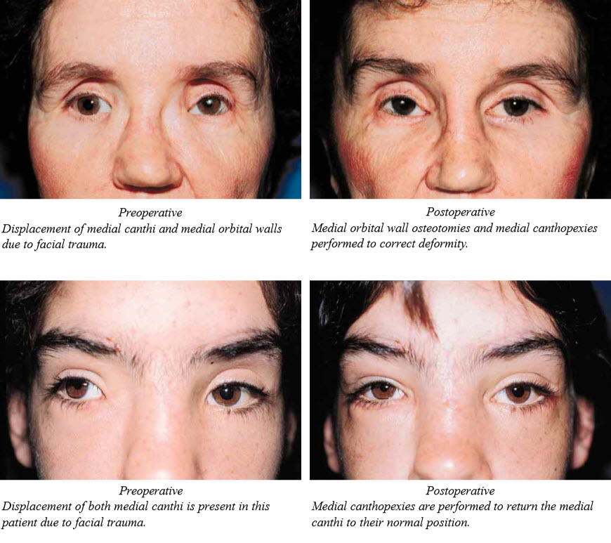
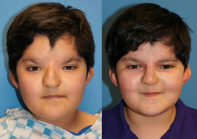
Many congenital and acquired deformities are associated with medial and lateral canthal displacement. Patients with blepharophimosis (telecanthus, epicanthal folds, ptosis), hypertelorism, Down’s Syndrome, craniofacial sysostosis, and acquired deformities may all have canthal deformities. Evaluation of the position and shape of the canthal area is a necessity in the planning of all orbital surgery. The contour and position of the canthi are important components in the aesthetic balance and symmetry of the face.
Canthal surgery essentially consists of repositioning the involved canthal tendon to the desired position and securing it to the bone. This would seem to be a simple, straight-forward procedure. However, because of the complex anatomy of the medial canthal area and the difficulty in obtaining normal, symmetrical soft tissue contours, this procedure is neither as easy nor as predictable as it would appear to be. Attention to surgical technique, soft tissue tension and contour, bone mobilization and position, and healing of the tendon to bone — all are important in the ultimate results of canthal surgery.


